Caudate nucleus
Deep within each half of the brain lies the caudate nucleus. The caudate nucleus caudate nucleus a pair of brain structures that make up part of the basal ganglia.
Federal government websites often end in. Before sharing sensitive information, make sure you're on a federal government site. The site is secure. NCBI Bookshelf. Margaret E. Driscoll ; Pradeep C. Bollu ; Prasanna Tadi.
Caudate nucleus
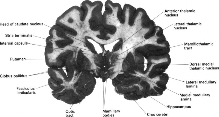
The caudate nucleus is one of the structures that make up the corpus striatum , which is a component of the basal ganglia in the human brain. The caudate is also one of the brain structures which compose the reward system and functions as part of the cortico — basal ganglia — thalamic loop. Together with the putamen , the caudate forms the dorsal striatum , which is considered a single functional structure; anatomically, it is separated by a large white matter tract, the internal capsule , so it is sometimes also referred to as two structures: the medial dorsal striatum the caudate and the lateral dorsal striatum the putamen. In this vein, the two are functionally distinct not as a result of structural differences, but merely due to the topographical distribution of function. The caudate nuclei are located near the center of the brain, sitting astride the thalamus. There is a caudate nucleus within each hemisphere of the brain. Individually, they resemble a C-shape structure with a wider "head" caput in Latin at the front, tapering to a "body" corpus and a "tail" cauda. Sometimes a part of the caudate nucleus is referred to as the "knee" genu. The head and body of the caudate nucleus form part of the floor of the anterior horn of the lateral ventricle. After the body travels briefly towards the back of the head, the tail curves back toward the anterior, forming the roof of the inferior horn of the lateral ventricle. This means that a coronal on a plane parallel to the face section that cuts through the tail will also cross the body and head of the caudate nucleus. The caudate is highly innervated by dopaminergic neurons that originate from the substantia nigra pars compacta SNc. The SNc is located in the midbrain and contains cell projections to the caudate and putamen , utilizing the neurotransmitter dopamine.
About Recent Edits Go ad-free. A study has suggested a link between Alzheimer's patients and the caudate nucleus.
At the time the article was last revised Yoshi Yu had no financial relationships to ineligible companies to disclose. Caudate nuclei are paired nuclei which along with the globus pallidus and putamen are referred to as the corpus striatum , and collectively make up the basal ganglia. The caudate nuclei have both motor and behavioral functions, in particular maintaining body and limb posture, as well as controlling approach-attachment behaviors, respectively 3. The caudate nucleus is located lateral to the lateral ventricles, with the head lateral to the frontal horn, and body lateral to the body of the lateral ventricle. The tail of the caudate nucleus terminates immediately above the temporal horn of the ventricle. It is bound laterally by the anterior crus of the internal capsule.
Federal government websites often end in. Before sharing sensitive information, make sure you're on a federal government site. The site is secure. NCBI Bookshelf. Margaret E. Driscoll ; Pradeep C. Bollu ; Prasanna Tadi. Authors Margaret E. Driscoll 1 ; Pradeep C.
Caudate nucleus
Our decisions often balance what we observe and what we desire. A prime candidate for implementing this complex balancing act is the basal ganglia pathway, but its roles have not yet been examined experimentally in detail. Here, we show that a major input station of the basal ganglia, the caudate nucleus, plays a causal role in integrating uncertain visual evidence and reward context to guide adaptive decision-making. In monkeys making saccadic decisions based on motion cues and asymmetric reward-choice associations, single caudate neurons encoded both sources of information.
Adaptador ac dc
Ultrasound Med Biol. ISBN How Well Do You Sleep? NCBI Bookshelf. The International Journal of Neuropsychopharmacology. Clear Turn Off Turn On. The caudate nucleus is not a common site of hemorrhage, but patients with caudate nucleus hemorrhage present with symptoms suggestive of subarachnoid hemorrhages such as headache, emesis, and nuchal rigidity. In perhaps the most illustrative case, a trilingual subject with a lesion to the caudate was observed. Loading Stack - 0 images remaining. The "motor release" observed as a result of this procedure indicates that the caudate nucleus inhibits the tendency for an animal to move forward without resistance. Like the lateral ventricles, the caudate is a C-shaped structure with a thick anterior portion called the head , which becomes narrower as it extends towards the back of the brain. The prognosis of caudate strokes has been considered good and benign, with majorities of individuals recovering and becoming independent. Follow NCBI. Prog Neurobiol.
It plays a critical role in various higher neurological functions. Each caudate nucleus is composed of a large anterior head, a body, and a thin tail that wraps anteriorly such that the caudate nucleus head and tail can be visible in the same coronal cut.
Deep brain stimulation in the ventral caudate nucleus is successful in the treatment of treatment-resistant obsessive-compulsive disorder and major depression, resulting in remission of depression and obsessive-compulsive symptoms around one year after implantation. Frontiers of Neurology and Neuroscience. The dentate gyrus and olfactory bulb are the two widely accepted sites of neurogenesis in adult mammals. Annu Rev Pathol. Efferent projections from the caudate nucleus travel to the hippocampus, globus pallidus, and thalamus. Review Connectivity patterns of thalamic nuclei implicated in dyskinesia. Volumetric variations in the caudate nucleus have been linked to many neurologic and psychiatric disorders, as outlined in the clinical significance section. Research has implicated caudate nucleus dysfunction in several pathologies, including Huntington and Parkinson disease, various forms of dementia, ADHD, bipolar disorder, obsessive-compulsive disorder, and schizophrenia. The caudate nucleus of those who speak multiple languages are larger than those who speak only one language, and the left caudate nucleus changes morphologically with multilingual expertise. Early symptoms are attributable to functions of the striatum and its cortical connections—namely control over movement, mood and higher cognitive function. Caudate nucleus as a component of networks controlling behavior.


I consider, that you are not right.
You are not right. Let's discuss it. Write to me in PM, we will talk.
I am sorry, that has interfered... I here recently. But this theme is very close to me. Write in PM.