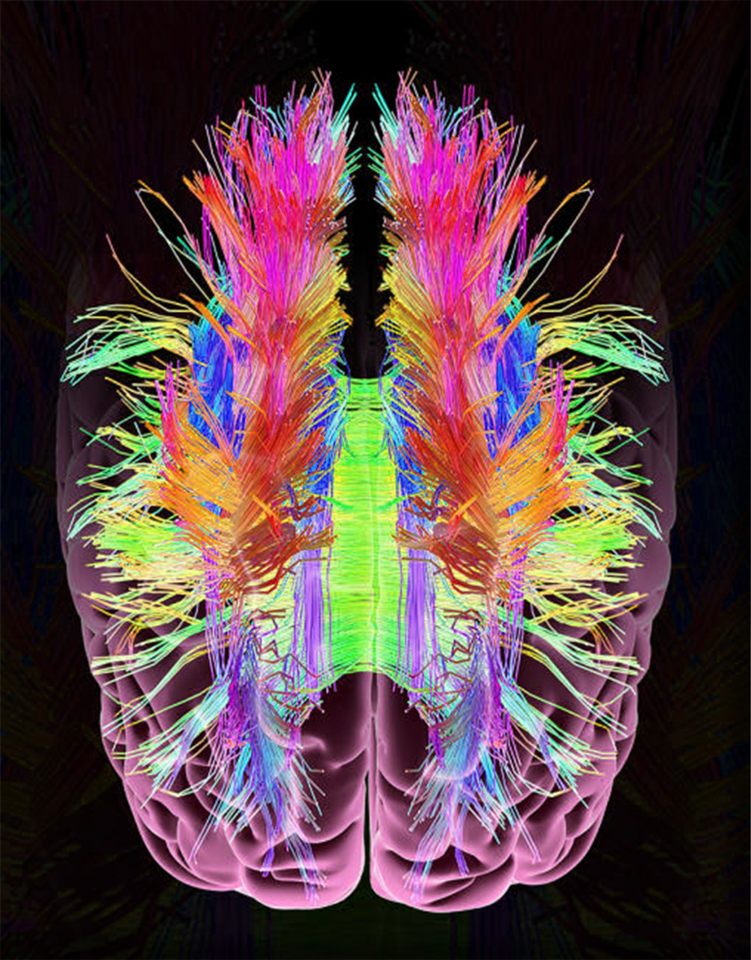
Diffusion tensor imaging
Functional MRI is a noninvasive diagnostic test that measures small changes in blood flow as a person performs tasks while in the MRI scanner. It detects the diffusion tensor imaging in action e.
At the time the article was last revised Rohit Sharma had no financial relationships to ineligible companies to disclose. Diffusion tensor imaging DTI is an MRI technique that uses anisotropic diffusion to estimate the axonal white matter organization of the brain. Fiber tractography FT is a 3D reconstruction technique to assess neural tracts using data collected by diffusion tensor imaging. Diffusion-weighted imaging DWI is based on the measurement of thermal Brownian motion of water molecules. Within cerebral white matter, water molecules tend to diffuse more freely along the direction of axonal fascicles rather than across them.
Diffusion tensor imaging
Diffusion-weighted magnetic resonance imaging DWI or DW-MRI is the use of specific MRI sequences as well as software that generates images from the resulting data that uses the diffusion of water molecules to generate contrast in MR images. Molecular diffusion in tissues is not random, but reflects interactions with many obstacles, such as macromolecules , fibers, and membranes. Water molecule diffusion patterns can therefore reveal microscopic details about tissue architecture, either normal or in a diseased state. A special kind of DWI, diffusion tensor imaging DTI , has been used extensively to map white matter tractography in the brain. In diffusion weighted imaging DWI , the intensity of each image element voxel reflects the best estimate of the rate of water diffusion at that location. Because the mobility of water is driven by thermal agitation and highly dependent on its cellular environment, the hypothesis behind DWI is that findings may indicate early pathologic change. A variant of diffusion weighted imaging, diffusion spectrum imaging DSI , [4] was used in deriving the Connectome data sets; DSI is a variant of diffusion-weighted imaging that is sensitive to intra-voxel heterogeneities in diffusion directions caused by crossing fiber tracts and thus allows more accurate mapping of axonal trajectories than other diffusion imaging approaches. Diffusion-weighted images are very useful to diagnose vascular strokes in the brain. It is also used more and more in the staging of non-small-cell lung cancer , where it is a serious candidate to replace positron emission tomography as the 'gold standard' for this type of disease. Diffusion tensor imaging is being developed for studying the diseases of the white matter of the brain as well as for studies of other body tissues see below. DWI is most applicable when the tissue of interest is dominated by isotropic water movement e.
Hybrid diffusion imaging HYDI. Fillard, P.
Diffusion tensor imaging DTI allows a live look into the microstructure of white matter in the brain and is an important complement to volumetric studies of specific structures such as the amygdala. DTI may be particularly informative for the study of autism because it has been speculated that white matter the connections between neurons defects may be even more pronounced than gray matter defects for affected individuals. Furthermore, in contrast to gray matter, white matter volume continues to increase across childhood and adolescence, thus allowing for analyses of growth curves and changes specific to the microstructure of axons. See papers by Courchesne, et al. Studies that utilize DTI technology generally describe two characteristics of white matter within a particular "voxel" in the brain:.
Although DTI and fiber tracking methods are proving valuable in the research realm for groups of subjects, I would caution their application for clinical diagnosis in individual patients. First, considerable variation of regional FA values exists among subjects as a function of age, sex, location, and MR technique. To reduce errors due to crossing, kissing, or branching fibers, a minimum of 20 diffusion directions should be obtained with 64 or more preferred. If FA values are compared to normal data bases, it is critical that multiple comparison e. Bonferroni corrections be performed to avoid spurious conclusions of statistical significance when none exist. Tractography results are even more variable than regional FA measurements and are highly dependent on the exact location of the initial seed points and tracking algorithms employed. An arbitrary starting point must be selected, whose exact location, size and orientation significantly affects results. Tractography algorithms differ in detail between vendors and arbitrary decisions must be made as to when tracking should stop.
Diffusion tensor imaging
The precise characterization of cerebral thrombi prior to an interventional procedure can ease the procedure and increase its success. This study investigates how well cerebral thrombi can be characterized by computed tomography CT , magnetic resonance MR and histology, and how parameters obtained by these methods correlate with each other as well as with the interventional procedure and clinical parameters. Cerebral thrombi of 25 patients diagnosed by CT with acute ischemic stroke were acquired by mechanical thrombectomy and, subsequently, scanned by a high spatial-resolution 3D MRI including T 1 -weighted imaging, apparent diffusion coefficient ADC , T 2 mapping and then finally analyzed by histology.
Xl public house salinas
This quantity is an assessment of the degree of restriction due to membranes and other effects and proves to be a sensitive measure of degenerative pathology in some neurological conditions. Aging 33, 21— A geometric comparison of diffusion anisotropy measures. The versatile nature of MRI is due to this capability of producing contrast related to the structure of tissues at the microscopic level. Nature Reviews. Burgel, U. Among most popular models are the biexponential model, which assumes the presence of 2 water pools in slow or intermediate exchange [17] [18] and the cumulant-expansion also called Kurtosis model, [19] [20] [21] which does not necessarily require the presence of 2 pools. A combined fMRI and DTI examination of functional language lateralization and arcuate fasciculus structure: effects of degree versus direction of hand preference. Diffusion imaging, white matter, and psychopathology. Barrick, T.
Thank you for visiting nature. You are using a browser version with limited support for CSS. To obtain the best experience, we recommend you use a more up to date browser or turn off compatibility mode in Internet Explorer.
Just Pretty Pictures? PMC About journal About journal. Frontiers in Surgery. The usage of 30 diffusion-encoded images orientations was found to be a good compromise between image quality and scanning time, since increasing the number of orientations didn't result in improved tensor orientation and MD estimates Jones, A review of diffusion tensor magnetic resonance imaging computational methods and software tools. A diffusion tensor imaging tractography atlas for virtual in vivo dissections. Different estimation methods may yield different results, therefore it is important to assure that the same package is used to estimate the tensors in an entire dataset Koay et al. Tensors can also be used to describe quantities that have directionality, such as mechanical force. Another commonly used measure that summarizes the total diffusivity is the Trace —which is the sum of the three eigenvalues,. The particular errors depend strongly on the tractography algorithm employed, and on the type of diffusion data used DTI versus higher-order models.


0 thoughts on “Diffusion tensor imaging”