Dorsal cochlear nucleus
Metrics details. The dorsal cochlear nucleus DCN is a region known to integrate somatosensory and auditory inputs and is identified as a potential key structure in the generation of phantom sound perception, especially noise-induced tinnitus. Yet, how altered homeostatic plasticity of the DCN induces and maintains the sensation of tinnitus is not dorsal cochlear nucleus. Mice were exposed to loud noise 9—11kHz, dorsal cochlear nucleus, 90dBSPL, 1h, followed by 2h of silenceand auditory brainstem responses ABRs and gap prepulse inhibition of acoustic startle GPIAS were recorded 2 days before and 2 weeks after noise exposure to identify animals with a significantly decreased inhibition of startle, indicating tinnitus but without permanent hearing loss.
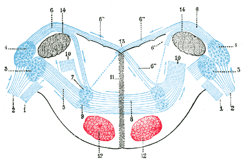
The dorsal cochlear nucleus DCN , also known as the " tuberculum acusticum " is a cortex-like structure on the dorso-lateral surface of the brainstem. Along with the ventral cochlear nucleus VCN , it forms the cochlear nucleus CN , where all auditory nerve fibers from the cochlea form their first synapses. The DCN differs from the ventral portion of the CN as it not only projects to the central nucleus a subdivision of the inferior colliculus CIC , but also receives efferent innervation from the auditory cortex , superior olivary complex and the inferior colliculus. The cytoarchitecture and neurochemistry of the DCN is similar to that of the cerebellum , an important concept in theories of DCN function. The pyramidal cells or giant cells are a major cell grouping of the DCN. These cells are the target of two different input systems. The first system arises from the auditory nerve, and carries acoustic information.
Dorsal cochlear nucleus
Federal government websites often end in. The site is secure. Author contributions: Z. The dorsal cochlear nucleus DCN is one of the first stations within the central auditory pathway where the basic computations underlying sound localization are initiated and heightened activity in the DCN may underlie central tinnitus. The neurotransmitter serotonin 5-hydroxytryptamine; 5-HT , is associated with many distinct behavioral or cognitive states, and serotonergic fibers are concentrated in the DCN. However, it remains unclear what is the function of this dense input. This excitatory effect results from an augmentation of hyperpolarization-activated cyclic nucleotide-gated channels I h or HCN channels. The serotonergic regulation of excitability is G-protein-dependent and involves cAMP and Src kinase signaling pathways. Moreover, optogenetic activation of serotonergic axon terminals increased excitability of fusiform cells. Our findings reveal that 5-HT exerts a potent influence on fusiform cells by altering their intrinsic properties, which may enhance the sensitivity of the DCN to sensory input. The serotonergic system modulates diverse physiological and behavioral functions, such as sleep, feeding, nociception, mood, and emotions Lucki, Serotonergic dysfunction has been implicated in a variety of psychiatric disorders, including depression, anxiety, schizophrenia, Parkinson's disease, and Alzheimer disease Meltzer et al.
Soc Neurosci Abs.
Federal government websites often end in. The site is secure. Tinnitus, the perception of a phantom sound, is a common consequence of damage to the auditory periphery. A major goal of tinnitus research is to find the loci of the neural changes that underlie the disorder. Crucial to this endeavor has been the development of an animal behavioral model of tinnitus, so that neural changes can be correlated with behavioral evidence of tinnitus.
Purpose: Eight lines of evidence implicating the dorsal cochlear nucleus DCN as a tinnitus contributing site are reviewed. We now expand the presentation of this model, elaborate on its essential details, and provide answers to commonly asked questions regarding its validity. Conclusions: Over the past decade, numerous studies have converged to support the hypothesis that the DCN may be an important brain center in the generation and modulation of tinnitus. Although other auditory centers have been similarly implicated, the DCN deserves special emphasis because, as a primary acoustic nucleus, it occupies a potentially pivotal position in the hierarchy of functional processes leading to the emergence of tinnitus percepts. Moreover, because a great deal is known about the underlying cellular categories and the details of synaptic circuitry within the DCN, this brain center offers a potentially powerful model for probing mechanisms underlying tinnitus. Abstract Purpose: Eight lines of evidence implicating the dorsal cochlear nucleus DCN as a tinnitus contributing site are reviewed. Publication types Review.
Dorsal cochlear nucleus
The dorsal cochlear nucleus DCN , also known as the " tuberculum acusticum " is a cortex-like structure on the dorso-lateral surface of the brainstem. Along with the ventral cochlear nucleus VCN , it forms the cochlear nucleus CN , where all auditory nerve fibers from the cochlea form their first synapses. The DCN differs from the ventral portion of the CN as it not only projects to the central nucleus a subdivision of the inferior colliculus CIC , but also receives efferent innervation from the auditory cortex , superior olivary complex and the inferior colliculus. The cytoarchitecture and neurochemistry of the DCN is similar to that of the cerebellum , an important concept in theories of DCN function. The pyramidal cells or giant cells are a major cell grouping of the DCN. These cells are the target of two different input systems.
Heart tonik faydaları
This would call into question the applicability of rodent models. Liu, S. However, evidence for these projections in mice, an increasingly important species in auditory neuroscience, is lacking, raising questions about the universality of such proposed functions. He found that the laminar organization and radial orientation of fusiform cells varied among species, with a very clear fusiform layer in tree shrews and to a lesser extent in bush babies and marmosets, whereas fusiform cells were randomly distributed in the gibbon. Figure 4. Inset: D: deep layer; F: fusiform cell layer; M: molecular layer. Hyperpolarizations of smaller amplitude in the postjunctional cell was taken as evidence of coupling. DCN unit activity was recorded in response to short sound pulses 3ms; 8—10, 9—11, 10—12, 12—14, and 14—16kHz filtered uniform white noise at different sound intensities 80—35dBSPL, 5dBSPL decreasing steps; presented at 10Hz. The pinna selectively amplifies frequencies, resulting in reduced sound energy at specific frequencies in certain regions of space. The trigeminal nerves are brightly labeled, as are axons in the spinal cord that likely originate from dorsal root ganglia. The literature is still ambiguous on how firing rate and tuning width are correlated in DCN units, since generalized inhibition by intracerebral injection of muscimol GABAA receptor agonist was shown to not affect tuning width in DCN units of anesthetized rats [ 67 ], while another study showed that bicuculline a GABA receptor antagonist increased tuning width while GABA and muscimol decreased tuning width [ 68 ]. The non-auditory domain receives mossy fiber input to granule cells, and is modified by Golgi and unipolar brush cells. The spectrum of behaviors influenced by serotonin. Dissection of brainstem. This coupling was present in mice up to 9 weeks postnatal.
Federal government websites often end in. The site is secure.
Sodium salicylate suppresses serotonin-induced enhancement of GABAergic spontaneous inhibitory postsynaptic currents in rat inferior colliculus in vitro. Characteristics of tinnitus and etiology of associated hearing loss: a study of patients. Relationship between auditory thresholds, central spontaneous activity, and hair cell loss after acoustic trauma. The glucose oxidase-DAB-nickel method in peroxidase histochemistry of the nervous system. The literature is still ambiguous on how firing rate and tuning width are correlated in DCN units, since generalized inhibition by intracerebral injection of muscimol GABAA receptor agonist was shown to not affect tuning width in DCN units of anesthetized rats [ 67 ], while another study showed that bicuculline a GABA receptor antagonist increased tuning width while GABA and muscimol decreased tuning width [ 68 ]. Shore, S. The rectangle shows the area in the larger image in B. December Learn how and when to remove this template message. Information in each nerve fibre is represented by the rate of action potentials as well as the particular timing of individual action potentials. Presumably, SSCs have the capacity to suppress excitatory synaptic signals to those dendrites in a region specific manner. Because I h time constants increase with depolarization Chen et al. Functional implications Given the importance of the DCN in auditory function and tinnitus pathophysiology, the serotonergic regulation of neuronal excitability of the primary output neurons may have important outcomes. Identifying the neurons involved in carrying these trigeminal signals to the auditory system will be crucial to understanding the neural mechanisms of somatic tinnitus. The rat In recent years, the rat has become a more popular experimental subject, especially in the development of an animal model of tinnitus Jastreboff and Sasaki, ; Lobarinas et al. Noise overexposure is known to alter firing properties of DCN cells [ 10 — 14 ], even after brief sound exposure at loud intensities [ 15 ].


I am sorry, that I interfere, I too would like to express the opinion.