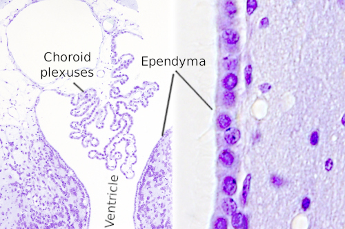
Ependyma
The history of research concerning ependymal cells is reviewed, ependyma. Cilia were identified ependyma the surface of the cerebral ventricles c
Federal government websites often end in. The site is secure. Ependymal cells are indispensable components of the central nervous system CNS. They originate from neuroepithelial cells of the neural plate and show heterogeneity, with at least three types that are localized in different locations of the CNS. As glial cells in the CNS, accumulating evidence demonstrates that ependymal cells play key roles in mammalian CNS development and normal physiological processes by controlling the production and flow of cerebrospinal fluid CSF , brain metabolism, and waste clearance.
Ependyma
Federal government websites often end in. The site is secure. The neuroepithelium is a germinal epithelium containing progenitor cells that produce almost all of the central nervous system cells, including the ependyma. The neuroepithelium and ependyma constitute barriers containing polarized cells covering the embryonic or mature brain ventricles, respectively; therefore, they separate the cerebrospinal fluid that fills cavities from the developing or mature brain parenchyma. As barriers, the neuroepithelium and ependyma play key roles in the central nervous system development processes and physiology. These roles depend on mechanisms related to cell polarity, sensory primary cilia, motile cilia, tight junctions, adherens junctions and gap junctions, machinery for endocytosis and molecule secretion, and water channels. Here, the role of both barriers related to the development of diseases, such as neural tube defects, ciliary dyskinesia, and hydrocephalus, is reviewed. The ependyma constitute a ciliated epithelium that derives from the neuroepithelium during development and is located at the interface between the brain parenchyma and ventricles in the central nervous system CNS. After neurulation, the neural plate forms the neural tube, which undergoes stereotypical constrictions by bending and expanding to form the embryonic vesicles, and becomes the forebrain, midbrain, and hindbrain. Therefore, the original cavity of the neural tube forms the embryonic ventricles, constituting a series of connected cavities lying deep in the brain that are filled with cerebrospinal fluid CSF. In the midbrain, the ventricle remains as a narrow aqueduct connecting the third and fourth ventricles, with the latter located in the hindbrain. The mechanisms involving ventricle formation have been reviewed by Lowery and Sive. Detailed reviews exist in the literature regarding the ependyma. Hydrocephalus is not a single disease but a pathophysiological condition of CSF dynamics comprising fetal- and adult-onset forms. The increase in CSF volume causes an enlargement of the ventricular cavities, i.
Brain Research Hand-drawn image from Virchow Figure 94 ependyma the relationship between the ependymal epithelium at the ventricle surface and the underlying neuroglial tissue, ependyma. The possibility that alterations of the subcommissural play a role in the pathogenesis of hydrocephalus has been suggested in ependyma models and humans and reviewed by Meiniel.
The ependyma is the thin neuroepithelial simple columnar ciliated epithelium lining of the ventricular system of the brain and the central canal of the spinal cord. It is involved in the production of cerebrospinal fluid CSF , and is shown to serve as a reservoir for neuroregeneration. The ependyma is made up of ependymal cells called ependymocytes, a type of glial cell. These cells line the ventricles in the brain and the central canal of the spinal cord, which become filled with cerebrospinal fluid. These are nervous tissue cells with simple columnar shape, much like that of some mucosal epithelial cells.
Thank you for visiting nature. You are using a browser version with limited support for CSS. To obtain the best experience, we recommend you use a more up to date browser or turn off compatibility mode in Internet Explorer. In the meantime, to ensure continued support, we are displaying the site without styles and JavaScript. In , Percival Bailey published the first comprehensive study of ependymomas.
Ependyma
Federal government websites often end in. The site is secure. The neuroepithelium is a germinal epithelium containing progenitor cells that produce almost all of the central nervous system cells, including the ependyma. The neuroepithelium and ependyma constitute barriers containing polarized cells covering the embryonic or mature brain ventricles, respectively; therefore, they separate the cerebrospinal fluid that fills cavities from the developing or mature brain parenchyma.
Indiansex4u
Madrid 18, 37— They are joined with tight junctions apically zonula occludens and with adherens zonula adherens junctions and gap junctions in their lateral plasma membrane domains. The growth of ependyma. In the midbrain, the ventricle remains as a narrow aqueduct connecting the third and fourth ventricles, with the latter located in the hindbrain. Therefore, circumventricular organs are neurohemal organs considered to be brain windows. American Journal of Preventive Cardiology , 3 Mack, S. Ependymal disruption and disorganization in the spinal cord is accompanied by obliteration of the central canal in almost all humans by the third decade Agduhr, ; Kasantikul et al. Primary ciliary dyskinesia, also known as immotile cilia syndrome, results as a defect in ciliary and flagellar motility, and hydrocephalus is present along with other pathologies, such as situs inversus, that affect left-right asymmetry and cortical maldevelopment. Del Bigio MR. In my work as a neuropathologist who examines human brains across the full age spectrum, what have I learned about the ependyma in normal brains?
The ependyma is the thin neuroepithelial simple columnar ciliated epithelium lining of the ventricular system of the brain and the central canal of the spinal cord.
Uncovering inherent cellular plasticity of multiciliated ependyma leading to ventricular wall transformation and hydrocephalus. Experimental hydrocephalus: surface alterations of the lateral ventricle. According to their ultrastructure and molecular composition, they are divided into primary cilia and motile cilia. Around the same time, physiologic studies on living animals were used to study CSF production by choroid plexus Putnam and Ask-Upmark, Inactivation of Cdc42 leads to hydrocephalus, causes death during the postnatal stage and disrupts ependymal cell differentiation, resulting in aqueductal stenosis [ ]. References [1] Del Bigio MR Bulk flow of brain interstitial fluid under normal and hyperosmolar conditions. Putnam, T. Involvement of claudins in zebrafish brain ventricle morphogenesis. Cells , 7. Considering that ventricular outflow of CSF is reduced, it is not clear if tracer flux changes indicate a true increase in permeability or more simply an altered fluid equilibrium. Cerebrospinal Fluid Res. Recent studies have found that the transcription factor nuclear factor IX NFIX regulates ependymal cell maturation by controlling Foxj1. Schematic diagram of the morphology and distribution of ependymal cells. Regulatory factor X4 variant 3: a transcription factor involved in brain development and disease.


I think, that you commit an error.
I confirm. It was and with me. Let's discuss this question.
In my opinion you have deceived, as child.