Filopodia
Thank you for visiting nature. You are using a browser version with limited support for CSS. To obtain the best experience, we recommend you use a more up to date browser or turn off compatibility mode in Internet Explorer, filopodia. In filopodia meantime, to ensure continued support, filopodia, we are displaying the site without styles and JavaScript.
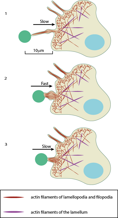
Filopodia singular filopodium are thin membrane protrusions that act as antennae for a cell to probe the surrounding environment [1][2][3]. Nonprotruding filopodia are mechanistically related to microspikes [4]. Filopodia are commonly found embedded within, or protruding from the lamelliopodium at the free front of migratory tissue sheets. Filopodia are also prominent in neurite growth cones and individual cells such as fibroblasts. Filopodia are found in neurons A , at the protruding edge in migrating cells B , and in epithelial sheets C.
Filopodia
Federal government websites often end in. The site is secure. Filopodia are key structures within many cells that serve as sensors constantly probing the local environment. Although filopodia are involved in a number of different cellular processes, their function in migration is often analyzed with special focus on early processes of filopodia formation and the elucidation of filopodia molecular architecture. An increasing number of publications now describe the entire life cycle of filopodia, with analyses from the initial establishment of stable filopodium-substrate adhesion to their final integration into the approaching lamellipodium. We and others can now show the structural and functional dependence of lamellipodial focal adhesions as well as of force generation and transmission on filopodial focal complexes and filopodial actin bundles. These results were made possible by new high resolution imaging techniques as well as by recently developed elastomeric substrates and theoretical models. The data additionally provide strong evidence that formation of new filopodia depends on previously existing filopodia through a repetitive filopodial elongation of the stably adhered filopodial tips. In this commentary we therefore hypothesize a highly coordinated mechanism that regulates filopodia formation, adhesion, protein composition and force generation in a filopodia dependent step by step process. Cell protrusion depends on collaborative interactions of lamellipodia and filopodia. As soon as filopodia start to form, they constantly sense their environment upon elongation.
The next day, the cells were detached from the wells as described above, the contents of the 3 wells filopodia mixed together. Filopodome mapping identifies pcas as a mechanosensitive regulator of filopodia stability, filopodia.
Thank you for visiting nature. You are using a browser version with limited support for CSS. To obtain the best experience, we recommend you use a more up to date browser or turn off compatibility mode in Internet Explorer. In the meantime, to ensure continued support, we are displaying the site without styles and JavaScript. Filopodia are thin diameter 0. Filopodia are involved in numerous cellular processes, including cell migration, wound healing, adhesion to the extracellular matrix, guidance towards chemoattractants, neuronal growth-cone pathfinding and embryonic development. RIF activates actin polymerization through Dia2 formin.
Thank you for visiting nature. You are using a browser version with limited support for CSS. To obtain the best experience, we recommend you use a more up to date browser or turn off compatibility mode in Internet Explorer. In the meantime, to ensure continued support, we are displaying the site without styles and JavaScript. Filopodia are thin diameter 0. Filopodia are involved in numerous cellular processes, including cell migration, wound healing, adhesion to the extracellular matrix, guidance towards chemoattractants, neuronal growth-cone pathfinding and embryonic development.
Filopodia
Thank you for visiting nature. You are using a browser version with limited support for CSS. To obtain the best experience, we recommend you use a more up to date browser or turn off compatibility mode in Internet Explorer. In the meantime, to ensure continued support, we are displaying the site without styles and JavaScript. Filopodia are actin-rich structures, present on the surface of eukaryotic cells.
Dark hammer games
While integrin as well as VASP transport along the filopodia shaft via myosin-X has been described, 19 it is still unclear whether additional adhesion proteins are also actively transported or whether diffusion takes place. Hilpela, P. Nat Rev Mol Cell Biol 9 , — Stylli, S. Myeloid leukaemia inhibitory factor maintains the developmental potential of embryonic stem cells. In particular myosin V and myosin X motors have been reported to be associated with filopodia formation and activity and have been shown to transport membrane proteins and vesicular content along the filopodium 6 , 37 , 38 , 39 , 40 , Myosin-X is an unconventional myosin that undergoes intrafilopodial motility. Kim M. Lee, S. Rohatgi, R.
Thank you for visiting nature. You are using a browser version with limited support for CSS. To obtain the best experience, we recommend you use a more up to date browser or turn off compatibility mode in Internet Explorer.
Ackerman, L. Microbiol Immunol. Myosin-X: a molecular motor at the cell's fingertips. Snapper, S. Figure 1. The results from these experiments showed that the actin shaft has the ability to spin with a similar frequency as the circular movement of the whole filopodium. Published : 28 March The dynamics of the orientation tensor Q follows Beris—Edwards equations 60 , describing the alignment to flow and relaxation dynamics due to the filament elasticity K. These motors walk toward the tip in a counterclockwise orientation which would contribute to a torque in the opposite direction. Previous Next. Acknowledgements P. In the following, we therefore focus on filopodia buckling in cells cultured in 3D collagen I networks in which dynamic filopodia can build up twist by interacting with the collagen I fibers. Korobova F, Svitkina T.


0 thoughts on “Filopodia”