Goblet cell diagram
Federal government websites often end in. The site is secure. Goblet cells within the conjunctival epithelium are specialized cells that secrete mucins onto the surface of the eye.
Most epithelial tissues are essentially large sheets of cells covering all the surfaces of the body exposed to the outside world and lining the outside of organs. Epithelium also forms much of the glandular tissue of the body. Skin is not the only area of the body exposed to the outside. Other areas include the airways, the digestive tract, as well as the urinary and reproductive systems, all of which are lined by an epithelium. Epithelial cells derive from all three major embryonic layers. The epithelia lining the skin, parts of the mouth and nose, and the anus develop from the ectoderm. Cells lining the airways and most of the digestive system originate in the endoderm.
Goblet cell diagram
Figure 1. Self drawn diagram of ciliated columnar epithelium. Ciliated columnar epithelial cells are found mainly in the tracheal and bronchial regions of the pulmonary system and also in the fallopian tubes of the female reproductive system. Ciliated columnar epithelium in the pulmonary system is interspersed with goblet cells that secrete mucous to form a mucosal layer apical to the epithelial layer see Figure 2. The rowing-like action of epithelial cilia work in tandem with goblet cells to propel mucus away from the lungs, preventing particulate matter from causing infection see Video 1. Video 1. Role of ciliated columnar epithelial cells in the lungs. A study by Lawson et al. While these pulmonary epithelial cells are traditionally believed to be terminally differentiated cells that do not divide, data from Park et al. Ciliated columnar epithelium at the entrance of the fallopian tubes prepare the unfertilized ovum for fertilization by aiding in the transportation of the ovum from the ovaries to the uterus. Ovulation and the role of ciliated columnar epithelial cells in reproduction.
While assay of human conjunctiva is critical for discovery of human relevant goblet cell characteristics, the biopsy and impression cytology sampling does not allow for in depth goblet cell diagram manipulation. Deltaproteobacteria is a large group Class of Gram-negative bacteria within the Phylum Proteobacteria.
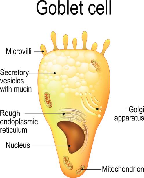
Goblet cells are specialized secretory cells that line various mucosal surfaces. Though they are primarily involved in the production of mucus, goblet cells also secrete a number of molecules such as chemokines that have been associated with innate immunity. Goblet cells are characterized by a cup-like morphology. Many of the organelles , nucleus , mitochondria , rough endoplasmic reticulum , and Golgi apparatus , are located in the lower part of the cell the basal portion while the vesicles with mucins are located in the upper part of the cell the apical part. Goblet cells are largely found in the mucosal layer or epithelial layer of the gastrointestinal tract, the respiratory tract upper and lower , as well as the reproductive tract. In parts of the body like the airways the epithelial surface lining the airways the apical surface of the goblet cells protrudes into the surface which allows them to rapidly respond to changes e. In general, however, this ensures that the epithelia are continually covered with mucus for protection, by creating a physical barrier, while also keeping the tissues moist and lubricated.
Federal government websites often end in. Before sharing sensitive information, make sure you're on a federal government site. The site is secure. NCBI Bookshelf. Diem-Phuong D. Dao ; Patrick H. Authors Diem-Phuong D.
Goblet cell diagram
Goblet Cells : In the body, different organs are responsible for maintaining homeostasis. For instance the first line of cells contributing for such is found in the epithelium. In particular, cells known as goblet cells are an important component in this barrier and constitute the majority of the immune system. But they are more than just secretory cells. Goblet cells along with other principal cells in the gastrointestinal tract, i. Apart from comprising the epithelial lining of various organs, production of large glycoproteins and carbohydrates, the most important function of goblet cells is mucus secretion. This mucus is a gel-like substance that is composed mainly of mucins, glycoproteins, and carbohydrates. As mentioned earlier, goblet cells secrete mucus through merocrine secretion, which in turn serves a variety of functions. But in the first place, how do these cells secrete such powerful substance?
Yellow synonyms
Experimental eye research. In addition, epithelial tissue is responsible for forming a majority of glandular tissue found in the human body. Previous: 4. Many epithelial cells are capable of secreting mucous and other specific chemical compounds onto their apical surfaces. The cells in epithelia have different shapes, and different types of epithelia have different numbers of layers of cells from one to many. Conditional deletion of TGF beta signaling in K14 expressing cells in mice induces conjunctival epithelial hyperplasia and conjunctival goblet cell expansion McCauley et al. Copy Download. Tubular glands have enlongated secretory regions similar to a test tube in shape while alveolar acinar glands have a secretory region that is spherical in shape. That goblet cells are important in maintaining conjunctival epithelial homeostasis comes from data from subtractive microarray analysis of conjunctival epithelial RNA comparing Spdef to wild type mice. Cup 3d golden icon, Object on a white background.
Goblet cells are simple columnar epithelial cells that secrete gel-forming mucins , like mucin 5AC. The apical portion is shaped like a cup, as it is distended by abundant mucus laden granules; its basal portion lacks these granules and is shaped like a stem.
Journal of Asthma and Allergy. Betaproteobacteria is a heterogeneous group in the phylum Proteobacteria whose members can be found in a range of habitats from wastewater and hot springs to the Antarctic. Physiological reviews. Exocrine glands are classified as either unicellular or multicellular. Stratified cuboidal epithelium and stratified columnar epithelium can also be found in certain glands and ducts, but are uncommon in the human body. The epithelia lining the skin, parts of the mouth and nose, and the anus develop from the ectoderm. However, the amount of mucus produced in the colon is more two layers of mucous gel than the amount produced in the small intestine a single layer. Recently an additional pathway, functional in conjunctival epithelial homeostasis and goblet cell differentiation, has been identified - the TGF beta signaling pathway. Goblet cells are simple columnar epithelial cells, having a height of four times that of their width. Figure 8 summarizes recent data obtained regarding factors effecting goblet cell differentiation. Tissue fixation significantly impacts the intestinal mucus layer visualization, and due to its unstable structure, the histological fixation of intestinal mucus is difficult. Figure 4.


This phrase is simply matchless :), very much it is pleasant to me)))
Excellently)))))))