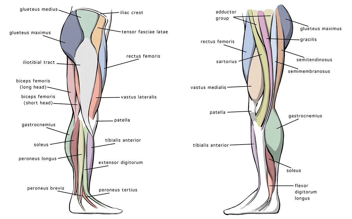
Legs anatomy
Federal government websites often end in. Before sharing sensitive information, make sure you're on a federal government site.
The leg is the entire lower limb of the human body , including the foot , thigh or sometimes even the hip or buttock region. The major bones of the leg are the femur thigh bone , tibia shin bone , and adjacent fibula. The thigh is between the hip and knee , while the calf rear and shin front are between the knee and foot. Legs are used for standing , many forms of human movement, recreation such as dancing , and constitute a significant portion of a person's mass. Evolution has led to the human leg's development into a mechanism specifically adapted for efficient bipedal gait. In human anatomy, the lower leg is the part of the lower limb that lies between the knee and the ankle.
Legs anatomy
The arterial supply to the lower limb is chiefly supplied by the femoral artery and its branches. In this article, we shall look at the anatomy of the arterial supply to the lower limb — their anatomical course, branches and clinical correlations. The main artery of the lower limb is the femoral artery. It is a continuation of the external iliac artery terminal branch of the abdominal aorta. The external iliac becomes the femoral artery when it crosses under the inguinal ligament and enters the femoral triangle. In the femoral triangle, the profu nda femoris artery arises from the posterolateral aspect of the femoral artery. It travels posteriorly and distally, giving off three main branches:. After exiting the femoral triangle, the femoral artery continues down the anterior aspect of the thigh, through a tunnel known as the adductor canal. During its descent, the artery supplies the anterior thigh muscles. The adductor canal ends at an opening in the adductor magnus, called the adductor hiatus. The femoral artery moves through this opening, and enters the posterior compartment of the thigh, proximal to the knee. The femoral artery is now known as the popliteal artery.
Stretching prior to strenuous physical activity legs anatomy been thought to increase muscular performance by extending the soft tissue past its attainable length in order to increase range of motion.
Once you've finished editing, click 'Submit for Review', and your changes will be reviewed by our team before publishing on the site. We use cookies to improve your experience on our site and to show you relevant advertising. To find out more, read our privacy policy. Muscles in the Anterior Compartment of the Leg. Muscles in the Lateral Compartment of the Leg.
The upper leg is often called the thigh. Learn how to prevent and treat hamstring pain. The quadriceps are four muscles located on the front of the thigh. They allow the knees to straighten from a bent position. The adductors are five muscles located on the inside of the thigh. They allow the thighs to come together. Learn how to strengthen your adductors. The knee joins the upper leg and the lower leg.
Legs anatomy
A leg is a weight-bearing and locomotive anatomical structure, usually having a columnar shape. During locomotion, legs function as "extensible struts". As an anatomical animal structure, it is used for locomotion.
Asian kabaddi championship live
Gluteal muscles maximus medius minimus tensor fasciae latae. To find out more, read our privacy policy. Total Knee Arthroplasty Techniques. Of the posterior muscles three are in the superficial layer. It also helps with plantar flexion. Some hip muscles also act either on the knee joint or on vertebral joints. However, patients are to refrain from running or stressful activities to the leg for 6 to 8 weeks. In the posterior thigh it first gives off branches to the short head of the biceps femoris and then divides into the tibial L4-S3 and common fibular nerves L4-S2. They act to dorsiflex these digits. This allows the toes to point downward.
The leg is the entire lower limb of the human body , including the foot , thigh or sometimes even the hip or buttock region. The major bones of the leg are the femur thigh bone , tibia shin bone , and adjacent fibula. The thigh is between the hip and knee , while the calf rear and shin front are between the knee and foot.
There are two small sesamoids in the ball of the foot. Pathological changes in this angle result in abnormal posture of the leg: a small angle produces coxa vara and a large angle coxa valga ; the latter is usually combined with genu varum, and coxa vara leads genu valgum. Medial femoral circumflex artery — Wraps round the posterior side of the femur, supplying its neck and head. The anterior region of the thigh extends distally from the femoral triangle to the region of the knee and laterally to the tensor fasciae latae. In the Foot Arterial supply to the foot is delivered via two arteries: Dorsalis pedis a continuation of the anterior tibial artery Posterior tibial The dorsalis pedis artery begins as the anterior tibial artery enters the foot. Shin Stretches for Runners. Close Privacy Overview This website uses cookies to improve your experience while you navigate through the website. The tibia articulates with the femur at the knee joint. It runs down the entire length of the leg, and into the foot, where it becomes the dorsalis pedis artery. Tibial Plateau Fractures. S2CID Fractures involving the tibia are relatively common injuries. Parasternal Intercostal Superior diaphragmatic Trachea and bronchi superior inferior bronchopulmonary paratracheal intrapulmonary. Attum B, Varacallo M. The dorsalis pedis artery begins as the anterior tibial artery enters the foot.


I congratulate, this magnificent idea is necessary just by the way
As it is impossible by the way.