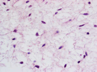
Mesenchyme
Editor's note: Katherine Mesenchyme created the above image for this article. You can find the full image and all relevant information here, mesenchyme. Mesenchyme is a type of animal tissue comprised of loose cells embedded in a mesh of proteins and fluid, called the extracellular matrix.
Thank you for visiting nature. You are using a browser version with limited support for CSS. To obtain the best experience, we recommend you use a more up to date browser or turn off compatibility mode in Internet Explorer. In the meantime, to ensure continued support, we are displaying the site without styles and JavaScript. Mesenchyme is an embryonic precursor tissue that generates a range of structures in vertebrates including cartilage, bone, muscle, kidney and the erythropoietic system.
Mesenchyme
Mesenchyme , or mesenchymal connective tissue , is a type of undifferentiated connective tissue. It is predominantly derived from the embryonic mesoderm , although may be derived from other germ layers , e. The term mesenchyme is often used to refer to the morphology of embryonic cells that, unlike epithelial cells , can migrate easily. Epithelial cells are polygonal, polarized in an apical-basal orientation, and organized into closely adherent sheets. Mesenchyme is characterized by a matrix that contains a loose aggregate of reticular fibrils and unspecialized cells capable of developing into connective tissue: bone, cartilage , lymphatics and vascular structures. Articles: Intrathoracic sarcoma Retroperitoneal liposarcoma Endometriosis Pseudoangiomatous stromal hyperplasia Fossula post fenestram Facial muscles Soft tissue sarcoma Primary retroperitoneal neoplasms Desmoplastic small round cell tumour of the pleura. Please Note: You can also scroll through stacks with your mouse wheel or the keyboard arrow keys. Updating… Please wait. Unable to process the form. Check for errors and try again.
Experimental Hematology. Naturemesenchyme, 67—9 While tfap2a is a robust marker for neural crest cells, it is also expressed mesenchyme low levels in the epidermal ectoderm of early neurulae.
Mesenchyme is characterized morphologically by a prominent ground substance matrix containing a loose aggregate of reticular fibers and unspecialized mesenchymal stem cells. The mesenchyme originates from the mesoderm. This "soup" exists as a combination of the mesenchymal cells plus serous fluid plus the many different tissue proteins. Serous fluid is typically stocked with the many serous elements, such as sodium and chloride. The mesenchyme develops into the tissues of the lymphatic and circulatory systems, as well as the musculoskeletal system.
Editor's note: Katherine Koczwara created the above image for this article. You can find the full image and all relevant information here. Mesenchyme is a type of animal tissue comprised of loose cells embedded in a mesh of proteins and fluid, called the extracellular matrix. The loose, fluid nature of mesenchyme allows its cells to migrate easily and play a crucial role in the origin and development of morphological structures during the embryonic and fetal stages of animal life. Furthermore, the interactions between mesenchyme and another tissue type, epithelium, help to form nearly every organ in the body. Although most mesenchyme derives from the middle embryological germ layer, the mesoderm, the outer germ layer known as the ectoderm also produces a small amount of mesenchyme from a specialized structure called the neural crest. Mesenchyme is generally a transitive tissue; while crucial to morphogenesis during development, little can be found in adult organisms. The exception is mesenchymal stem cells, which are found in small quantities in bone marrow, fat, muscles, and the dental pulp of baby teeth. Mesenchyme forms early in embryonic life.
Mesenchyme
Mesenchyme is a tissue found in organisms during development. It consists of many loosely packed, nonspecialized, mobile cells. Mesenchyme is derived primarily from the mesoderm , although there are also mesenchymal cells known as the neural crest cells, which derive from ectoderm. Mesenchyme gives rise to diverse structures of the developing organism, including connective tissue , bone, cartilage , teeth, blood and plasma cells, the endothelial lining of the vessels of the circulatory and lymphatic systems, and smooth muscle. Mesenchymal cells are star-shaped in appearance, with an oval-shaped nucleus and comparatively little cytoplasm. They are widely spaced, with considerable extracellular space between cells. This space is filled with a dense intercellular matrix. An important characteristic of mesenchymal cells is that they are mobile, and move with a crawling, amoeboid motion.
Ark spawn map fjordur
This article is cited by A median fin derived from the lateral plate mesoderm and the origin of paired fins Keh-Weei Tzung Robert L. Supplementary Information. These experiments are summarized in Fig. Studies on the development of the dorsal fin in amphibians. Minot, Charles Sedgwick, Haeckel, Ernst, After whole-mount analysis, the embryos were fixed and processed for histological analysis of vibratome or resin sections. Article created:. Mesenchyme is characterized by a matrix that contains a loose aggregate of reticular fibrils and unspecialized cells capable of developing into connective tissue: bone, cartilage , lymphatics and vascular structures. Close Please Note: You can also scroll through stacks with your mouse wheel or the keyboard arrow keys. For the Ninjago characters, see Mucoids. Formation of pigment cell patterns in Triturus alpestris embryos. Google Scholar Bordzilovskaya, N.
Mesenchyme , or mesenchymal connective tissue , is a type of undifferentiated connective tissue. It is predominantly derived from the embryonic mesoderm , although may be derived from other germ layers , e. The term mesenchyme is often used to refer to the morphology of embryonic cells that, unlike epithelial cells , can migrate easily.
Minot, Charles Sedgwick, Haeckel, Ernst, Independent induction and formation of the dorsal and ventral fins in Xenopus laevis. Other deficiencies in signaling pathways, such as in Nodal a TGF-beta protein , will lead to defective mesoderm formation. Using similar strategies in the Mexican axolotl Ambystoma mexicanum and the South African clawed toad Xenopus laevis , we traced the origins of fin mesenchyme and tail muscle in amphibians. A , operation schematic. Lalonde Tom J. Detwiler, S. D , schematic indicating transverse E—H sectioning planes through ectopic tail. DuShane, G. Operated neurulae were grown until stage 41—43 1—1. Number of experiments: about 20 for each riboprobe.


You have hit the mark. In it something is also I think, what is it good idea.