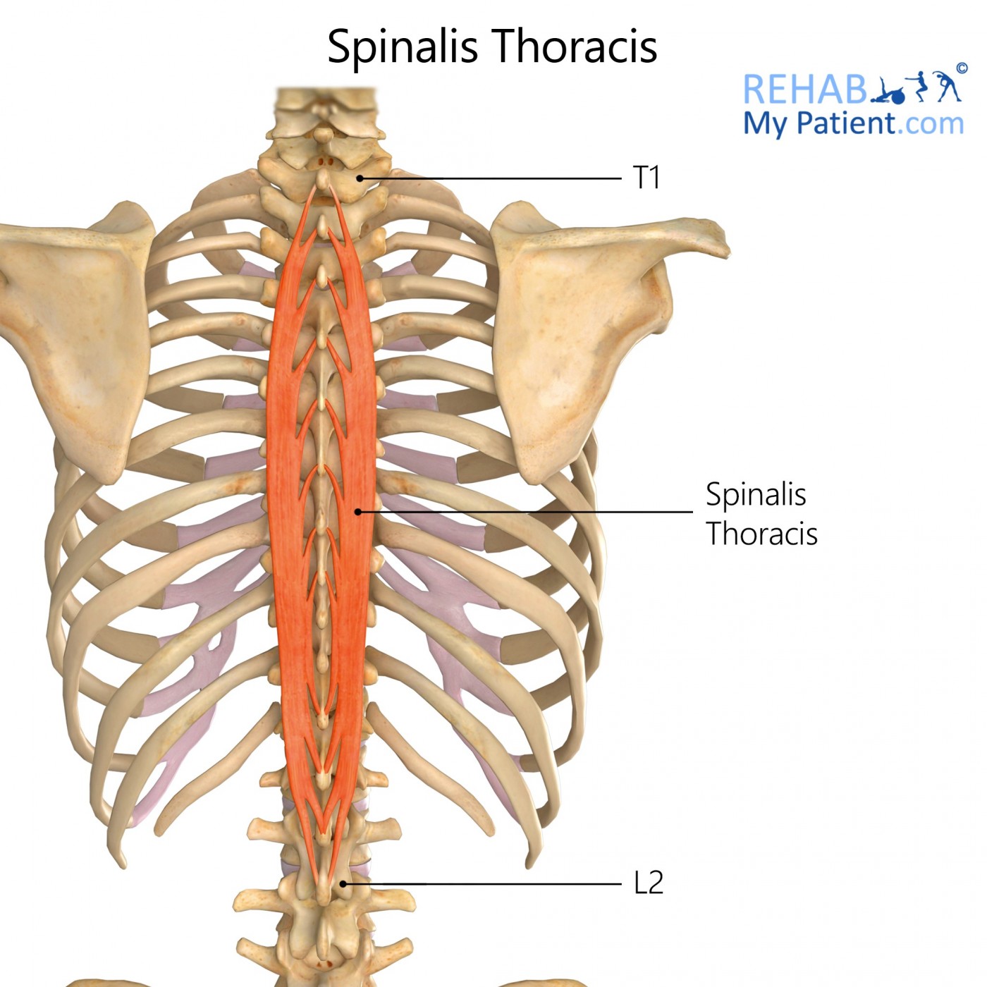
Spinalis origin and insertion
The spinalis is a deep muscle of the back.
The spinalis muscle is situated in the middle and upper back as well as the neck, running parallel to the spine. It plays an important role in extending the back and neck, while also aiding in lateral flexion movements. The spinalis muscle is a member of the erector spinae muscle group. The erector spinae muscles consist of the spinalis, iliocostalis , and the longissimus. The erector spinae muscles are deep muscles of the back which run in a vertical direction, parallel to the vertebral column.
Spinalis origin and insertion
The spinalis is a portion of the erector spinae , a bundle of muscles and tendons , located nearest to the spine. It is divided into three parts: Spinalis dorsi, spinalis cervicis, and spinalis capitis. Spinalis dorsi, the medial continuation of the sacrospinalis , is scarcely separable as a distinct muscle. It is situated at the medial side of the longissimus dorsi , and is intimately blended with it; it arises by three or four tendons from the spinous processes of the first two lumbar and the last two thoracic vertebrae : these, uniting, form a small muscle which is inserted by separate tendons into the spinous processes of the upper thoracic vertebrae, the number varying from four to eight. It is intimately united with the semispinalis dorsi , situated beneath it. Spinalis cervicis, or spinalis colli, is an inconstant muscle, which arises from the lower part of the nuchal ligament , the spinous process of the seventh cervical, and sometimes from the spinous processes of the first and second thoracic vertebrae , and is inserted into the spinous process of the axis , and occasionally into the spinous processes of the two cervical vertebrae below it. Spinalis capitis biventer cervicis is usually inseparably connected with the semispinalis capitis. Spinalis capitis is not well characterized in modern anatomy textbooks and atlases, and is often omitted from anatomical illustration. However, it can be identified as fibers that extend from the spinous processes of TV1 and CV7 to the cranium, often blending with semispinalis capitis [ citation needed ]. This article incorporates text in the public domain from page of the 20th edition of Gray's Anatomy Contents move to sidebar hide.
The spinalis is highlighted in yellow, the longissimus in purple, and the iliocostalis in green. Contents move to sidebar hide.
The spinalis muscle is the most medial of the erector spinae group of muscles, and is lateral to the multifidus group. The spinalis detaches from medial side of the longissimus thoracis and travels forward near thoracic vertebral spinous processes to cervical vertebral spinous processes. It may be divided into two parts:. In the pig and the horse, the spinalis muscle forms a common muscle belly, therefore sometimes termed as "spinalis thoracic et cervicis thoracic and cervical spinal muscle ", whereas in ruminants and carnivores, the thoracic and cervical spinalis muscles receive additional muscular strands from the the mamillary and transverse processes of some vertebrae, and the fibers of the spinlais musccles are closely related to and often difficult to separate form the semispinalis muscle. Therefore, some authors use the compound name "thoracic and cervical spinal and semispinal muscle" to describe this muscular complex. Origin: extends across the spinous processes of one or more thoracic vertebrae, and sometimes last cervical vertebra.
The spinalis is a deep muscle of the back. It is the smallest of the muscle columns within the erector spinae complex, and can be divided into the three parts — thoracic, cervicis and capitis although the cervicis part is absent in some individuals. It is the smallest of the muscle columns within the erector spinae complex, and can be divided into the three parts - thoracic, cervicis and capitis although the cervicis part is absent in some individuals. Once you've finished editing, click 'Submit for Review', and your changes will be reviewed by our team before publishing on the site. We use cookies to improve your experience on our site and to show you relevant advertising. To find out more, read our privacy policy. Spinalis Home Encyclopaedia S Spinalis. Not yet rated. Attachments: Arises from the lower thoracic and lumbar vertebrae, sacrum, posterior aspect of the iliac crest, and the sacroiliac and supraspinous ligament. It attaches to the spinous processes of C2, T1-T8 and the occipital bone of the skull.
Spinalis origin and insertion
Search site Search Search. Go back to previous article. Sign in. Eye Muscle Action Origin Insertion levator palpebrae superioris elevating and retracting the upper eyelid sphenoid bone upper eyelid inferior oblique looking up and laterally eye roll maxilla bone eyeball inferior, lateral inferior rectus looking down depression sphenoid bone eyeball inferior, medial lateral rectus looking laterally abduction sphenoid bone eyeball lateral, anterior medial rectus looking medially adduction sphenoid bone eyeball medial superior oblique looking down and laterally eye roll sphenoid bone eyeball superior, lateral superior rectus looking up elevation sphenoid bone eyeball superior, anterior. Thorax Muscle Action Origin Insertion diaphragm increasing thoracic volume for inhalation sternum, ribs, lumbar vertebrae central tendinous sheet external intercostals elevating ribs inferior aspects of ribs superior aspects of ribs innermost intercostals adducting the ribs, decreasing thoracic volume for exhalation inferior aspects of ribs superior aspects of ribs internal intercostals depressing ribs superior aspects of ribs inferior aspects of ribs pectoralis major flexing, adducting, and medially rotating the arm at the shoulder sternum, clavicle humerus pectoralis minor elevating the ribs, moving scapula anterior and inferior, protracting and elevating the shoulder ribs scapula serratus anterior moving and fixing scapula anteriorly, protracting the shoulder ribs scapula. Abdomen Muscle Action Origin Insertion external oblique flexing vertebral column, rotating vertebral column, compressing the abdomen lower ribs ilium, pubis, linea alba internal oblique flexing vertebral column, rotating vertebral column, compressing the abdomen lumbar vertebrae, ilium pubis, linea alba, lower ribs, sternum rectus abdominis flexing vertebral column, compressing the abdomen pubis lower ribs, sternum transversus abdominis compressing the abdomen lower ribs, ilium, lumbar vertebrae pubis, linea alba. Rotator Cuff Muscle Action Origin Insertion infraspinatus laterally rotating the shoulder scapula humerus subscapularis medially rotating the shoulder, stabilizes shoulder joint scapula humerus supraspinatus abducting the shoulder, stabilizes shoulder joint scapula humerus teres minor laterally rotating the shoulder scapula humerus. Arm Shoulder to Elbow Muscle Action Origin Insertion biceps brachii flexing the arm at the elbow scapula radius brachialis flexing the arm at the elbow humerus ulna coracobrachialis flexing and adducting the arm scapula humerus deltoid abducting the arm at the shoulder, flexing and extending arm at the shoulder clavicle, scapula humerus triceps brachii extending the arm at the elbow humerus, scapula ulna. Attributions "Anatomy and Physiology" by J.
Derivative using first principle
See more. It is situated at the medial side of the longissimus dorsi , and is intimately blended with it; it arises by three or four tendons from the spinous processes of the first two lumbar and the last two thoracic vertebrae : these, uniting, form a small muscle which is inserted by separate tendons into the spinous processes of the upper thoracic vertebrae, the number varying from four to eight. Menu Sign in. This article incorporates text in the public domain from page of the 20th edition of Gray's Anatomy The origins are highlighted in red, while the insertions are colored blue. Enter at least three characters in the search field. The spinalis capitis muscle is absent entirely in Spinalis cervicis, or spinalis colli, is an inconstant muscle, which arises from the lower part of the nuchal ligament , the spinous process of the seventh cervical, and sometimes from the spinous processes of the first and second thoracic vertebrae , and is inserted into the spinous process of the axis , and occasionally into the spinous processes of the two cervical vertebrae below it. Rectus abdominis muscle. To find out more, read our privacy policy.
The spinalis Latin: musculus spinalis is one of the muscles forming the erector spinae - a muscle complex consisting of several smaller intrinsic deep back muscle groups that all together form the intermediate layer of the deep back muscles. The other two groups are the longissimus and iliocostalis muscles.
It plays an important role in extending the back and neck, while also aiding in lateral flexion movements. Some of them require your consent. The thoracis section spans from L2 to T2 and affects the vertebrae in this area. Spinalis capitis biventer cervicis is usually inseparably connected with the semispinalis capitis. Thoracis: Spinous process of upper thoracic vertebrae. Muscles of the thorax and back. Recent searches. Unilateral contraction refers to the contraction of one side, while bilateral contraction refers to the contraction of both sides together [7]. Non-necessary Non-necessary. Once you've finished editing, click 'Submit for Review', and your changes will be reviewed by our team before publishing on the site.


It is interesting. Tell to me, please - where I can find more information on this question?