Transducin
Federal government websites often end in. The site is secure. To elucidate the determinants of G T coupling and activation, transducin, we obtained cryo-EM structures transducin a fully functional, light-activated Rho-G T complex in the presence and absence of a G protein-stabilizing nanobody, transducin.
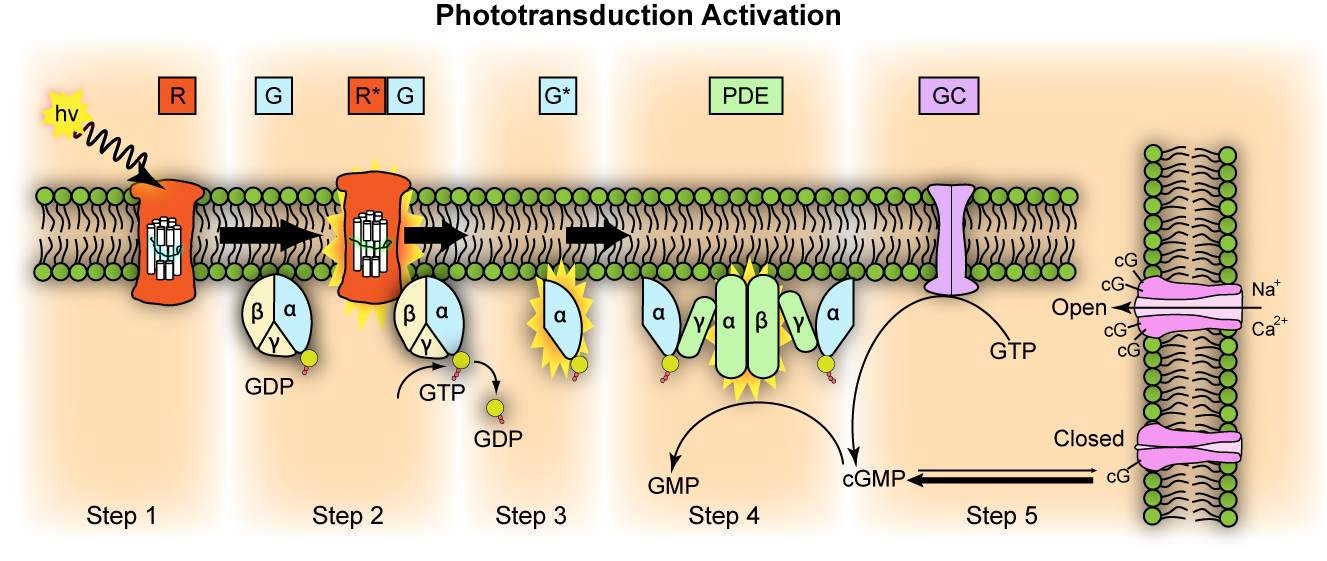
Transducin G t is a protein naturally expressed in vertebrate retina rods and cones and it is very important in vertebrate phototransduction. Light leads to conformational changes in rhodopsin , which in turn leads to the activation of transducin. Transducin activates phosphodiesterase , which results in the breakdown of cyclic guanosine monophosphate cGMP. The intensity of the flash response is directly proportional to the number of transducin activated. Transducin is activated by metarhodopsin II , a conformational change in rhodopsin caused by the absorption of a photon by the rhodopsin moiety retinal. Isomerization causes a change in the opsin to become metarhodopsin II.
Transducin
Transducin mediates signal transduction in a classical G protein-coupled receptor GPCR phototransduction cascade. Interactions of transducin with the receptor and the effector molecules had been extensively investigated and are currently defined at the atomic level. Protein-protein interactions underlying this modulation are largely unknown. We generated a mouse model with conditional knockout of Ric-8A in rods in order to begin defining the functional roles of the protein in rod photoreceptors and the retina. Traditionally, studies of transducin focused on its structure and mechanisms underlying this signaling cascade. Phototransduction takes place in a specialized ciliary compartment of photoreceptor cells called the outer segment OS. The remarkable molecular level of insight into these interactions has been recently elevated with solutions of the cryo-EM structures of transducin complexed with rhodopsin and PDE6 Gao et al. Nevertheless, several important aspects of transducin biology, including its folding, trafficking, and roles outside the phototransduction cascade remain largely obscure. Analyses of mouse models with impaired transducin translocation also support an important role of the phenomenon in neuroprotection of rods, presumably by reducing the metabolic stress associated with the constitutive phototransduction reactions under light conditions saturating responsiveness of rods Fain, ; Peng et al. UNC is a mammalian ortholog of C. Truncation mutation in UNC is linked to cone-rod dystrophy in human patients Kobayashi et al. Owing its lipid-binding specificity, UNC is generally viewed as a carrier protein for myristoylated cargo, with a preference for cargo proteins targeted to primary cilia via an ARL3-dependent mechanism Ismail et al.
Sulfhydryl-reactive, cleavable, transducin, and radioiodinatable benzophenone photoprobes for transducin of protein-protein interaction. Dynamics of cyclic GMP synthesis in retinal rods.
Federal government websites often end in. The site is secure. Transducin is a prototypic heterotrimeric G-protein mediating visual signaling in vertebrate photoreceptor cells. Heterotrimeric G-proteins have been long recognized to mediate a vast number of intracellular signaling pathways; however, the cellular mechanisms responsible for their assembly and intracellular targeting remain far from understood for review, see Marrari et al. Transducin or G t is one of the best studied G-proteins. It mediates phototransduction between the light-activated visual pigment rhodopsin and the effector enzyme cGMP phosphodiesterase PDE in retinal rods [for review, see Burns and Baylor , Fain et al. The rate of transducin activation, which is a key determinant in setting the photoreceptor's sensitivity to light Pugh and Lamb, , depends on transducin concentration in photoreceptor outer segments Sokolov et al.
Federal government websites often end in. The site is secure. Transducin is a prototypic heterotrimeric G-protein mediating visual signaling in vertebrate photoreceptor cells. Heterotrimeric G-proteins have been long recognized to mediate a vast number of intracellular signaling pathways; however, the cellular mechanisms responsible for their assembly and intracellular targeting remain far from understood for review, see Marrari et al. Transducin or G t is one of the best studied G-proteins. It mediates phototransduction between the light-activated visual pigment rhodopsin and the effector enzyme cGMP phosphodiesterase PDE in retinal rods [for review, see Burns and Baylor , Fain et al. The rate of transducin activation, which is a key determinant in setting the photoreceptor's sensitivity to light Pugh and Lamb, , depends on transducin concentration in photoreceptor outer segments Sokolov et al. However, the outer segment content of transducin changes during the normal diurnal cycle as a result of its reversible light-driven translocation from the outer segment to other cellular compartments for review, see Calvert et al. This phenomenon is thought to contribute to photoreceptor light adaptation by reducing transducin activation rate at bright light Sokolov et al.
Transducin
Transducin G t is a protein naturally expressed in vertebrate retina rods and cones and it is very important in vertebrate phototransduction. Light leads to conformational changes in rhodopsin , which in turn leads to the activation of transducin. Transducin activates phosphodiesterase , which results in the breakdown of cyclic guanosine monophosphate cGMP. The intensity of the flash response is directly proportional to the number of transducin activated. Transducin is activated by metarhodopsin II , a conformational change in rhodopsin caused by the absorption of a photon by the rhodopsin moiety retinal. Isomerization causes a change in the opsin to become metarhodopsin II. Decrease in cGMP concentration leads to decreased opening of cation channels and subsequently hyperpolarization of the membrane potential. This process is accelerated by a complex containing an RGS Regulator of G-protein Signaling -protein and the gamma-subunit of the effector, cyclic GMP phosphodiesterase. The amino terminal might be anchored or in close proximity to the carboxyl terminal for activation of the transducin molecule by rhodopsin.
American flooring niles
Further information and requests for reasorces and reagents should be directed to and will be fulfilled by the Lead Contact, Georgios Skiniotis moc. Figure 4. Transmission electron microscopy of rod outer segments from 0. Subunit dissociation and diffusion determine the subcellular localization of rod and cone transducins. Publish with us For authors Language editing services Submit manuscript. Subjects Enzyme mechanisms Retina. Pugh, E. The manuscript will undergo copyediting, typesetting, and review of the resulting proof before it is published in its final citable form. Skiba, and V. The role of mass transport limitation and surface heterogeneity in the biophysical characterization of macromolecular binding processes by SPR biosensing. Artemyev, N.
Federal government websites often end in. Before sharing sensitive information, make sure you're on a federal government site. The site is secure.
Full PDE activity was determined after treatment with trypsin trypsin-treated. Sci 28 , 13— J Physiol Lond ; — In that complex, the ICL2 helix exposes nonpolar residues at identical positions M Slepak, V. Phototransduction in mouse rods and cones. Structure of the G protein chaperone and guanine nucleotide exchange factor Ric-8A bound to Galphai1. Makino, E. Figure 4. Published : 10 May


Prompt to me please where I can read about it?